

Links to other sites are provided for information only - they do not constitute endorsements of those other sites. Macrocalcifications are typically benign and usually don’t need follow-up imaging. They’re the most common type of calcification found in breast tissue. Macrocalcifications appear as large white spots randomly scattered throughout your breasts. Calcifications in a noncancerous growth called a. There are two types of breast calcifications. Prior radiation therapy for breast cancer.
Breast calcification clusters skin#
Skin calcifications caused by dermatitis (a rash) or metallic residues from powder, deodorants or ointments. A licensed physician should be consulted for diagnosis and treatment of any and all medical conditions. Calcifications associated with a dilated milk duct.
Breast calcification clusters how to#
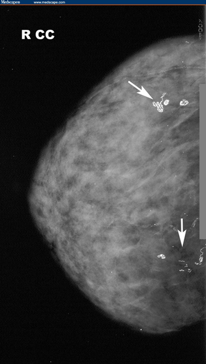
The information provided herein should not be used during any medical emergency or for the diagnosis or treatment of any medical condition. The appearance of clustered calcifications is one of the important signs of breast cancer, and thus how to classify clustered calcifications comes to be a key. This site complies with the HONcode standard for trustworthy health information: verify here. Learn more about A.D.A.M.'s editorial policy editorial process and privacy policy. is among the first to achieve this important distinction for online health information and services. follows rigorous standards of quality and accountability. is accredited by URAC, for Health Content Provider (URAC's accreditation program is an independent audit to verify that A.D.A.M. Most women who have suspicious calcifications do not have cancer.Ī.D.A.M., Inc. The purpose of the biopsy is to find out if the calcifications are benign (not cancer) or malignant (cancer). This is a needle biopsy that uses a type of mammogram machine to help find the calcifications. Your provider will recommend a stereotactic core biopsy. Most women will need to have a follow-up mammogram in 6 months.Ĭalcifications that are irregular in size or shape or are tightly clustered together, are called suspicious calcifications. In some cases, calcifications that are slightly abnormal but do not look like a problem (such as cancer) are also called benign. But, your health care provider may recommend that you get a mammogram each year. When microcalcifications are present on a mammogram, the doctor (a radiologist) may ask for a larger view so the areas can be examined more closely.Ĭalcifications that do not appear to be a problem are called benign. However, these areas may need to be checked more closely if they have a certain appearance on the mammogram. Microcalcifications are tiny calcium specks seen on a mammogram. They are most likely not related to cancer. They look like small white dots on the mammogram. Large, rounded calcifications (macrocalcifications) are common in women over age 50.


 0 kommentar(er)
